



Catheter Ablation
A detailed guide to catheter ablation for heart rhythm disorders, including mapping, preparation, risks, recovery and follow-up.
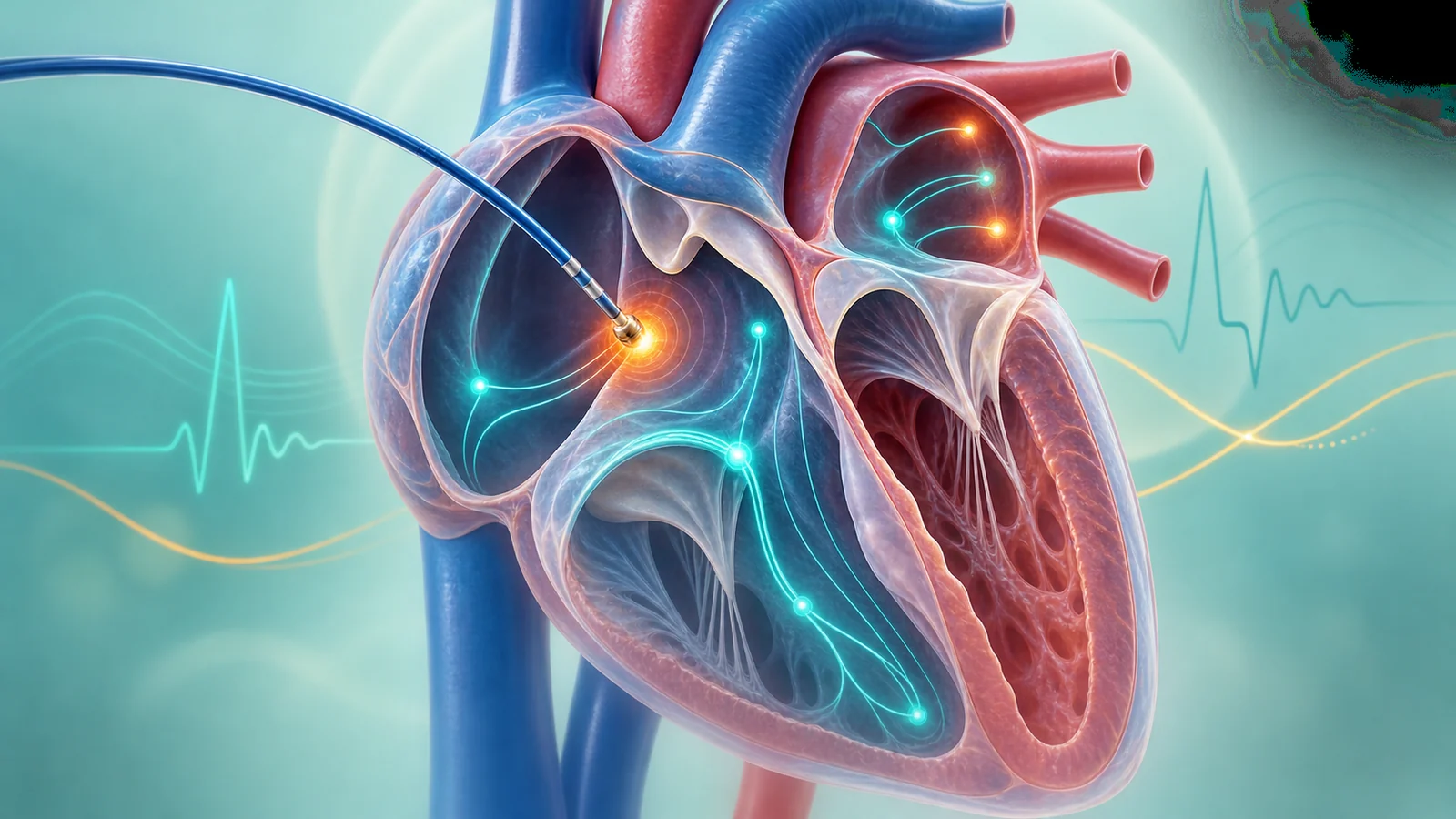
Catheter ablation is a minimally invasive treatment for selected heart rhythm disorders. Thin catheters are introduced through blood vessels and guided to the heart. Electrical recordings and three-dimensional mapping identify the tissue responsible for the abnormal rhythm. Energy is then delivered to modify a small, targeted area.
Which Rhythm Disorders Can Be Treated?
Ablation may be considered for supraventricular tachycardia, accessory pathways, typical atrial flutter, atrial fibrillation, selected atrial tachycardias, frequent premature ventricular beats and ventricular tachycardia. Expected success, procedure complexity and risk vary substantially between rhythm types. Symptoms, rhythm documentation, structural heart disease, medicine response and patient preference guide the decision.
Before the Procedure
An ECG or rhythm recording must establish the diagnosis. Echocardiography, blood tests and sometimes CT or MRI may be required. Instructions about anticoagulants, antiarrhythmic medicines, fasting and anaesthesia are individualised. Medicines should never be stopped without specific advice.
Mapping and Ablation
During an electrophysiology study, catheters record electrical activity and may stimulate the heart to reproduce the rhythm. Three-dimensional electroanatomical mapping can reduce reliance on fluoroscopy and improve localisation. Radiofrequency energy heats tissue, while cryoablation cools it. The chosen technique depends on the rhythm and anatomy.
Atrial fibrillation ablation most often targets electrical conduction around the pulmonary veins. More extensive ablation is not automatically beneficial for every patient. In complex ventricular arrhythmias, mapping may involve the inner or occasionally outer surface of the heart.
Benefits, Risks and Expectations
Ablation can reduce or eliminate episodes and improve quality of life, but recurrence is possible. Some patients require repeat treatment. Risks include bleeding or vascular injury, fluid around the heart, stroke, damage to the normal conduction system, narrowing of a pulmonary vein, oesophageal injury in atrial fibrillation procedures and, rarely, death. The risk profile must be explained for the specific procedure.
Recovery and Follow-up
The access sites, heart rhythm and vital signs are monitored after the procedure. Temporary palpitations can occur during healing and do not always indicate failure. Follow-up may include ECG, Holter or longer rhythm monitoring. Anticoagulation after atrial fibrillation ablation is guided mainly by stroke risk and should not be stopped because symptoms have disappeared.
When to Seek Emergency Help
Fever, difficulty swallowing, neurological symptoms or progressive groin swelling after an atrial fibrillation procedure also require prompt medical advice.
Frequently Asked Questions
Is ablation open-heart surgery?
No. It is usually performed through vascular catheters, without opening the chest.
Is general anaesthesia always required?
No. Sedation or general anaesthesia is selected according to the rhythm, procedure and patient.
Can arrhythmia recur?
Yes. Recurrence depends on rhythm type and clinical factors; repeat evaluation or ablation may sometimes be appropriate.